

Tissue Iron Detection
- Detect iron in tissue samples
- Standard samples for quantifying the content of Fe2+ and Fe3+ are included
- A convenient calculation sheet for calculating iron content is provided
Description
Iron is one of the most important metallic elements in living organisms. In recent years, an iron-dependent mode of cell death, ferroptosis, has emerged, and research on intracellular iron ions has attracted increased attention.
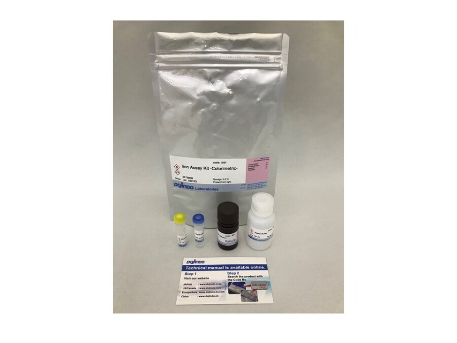
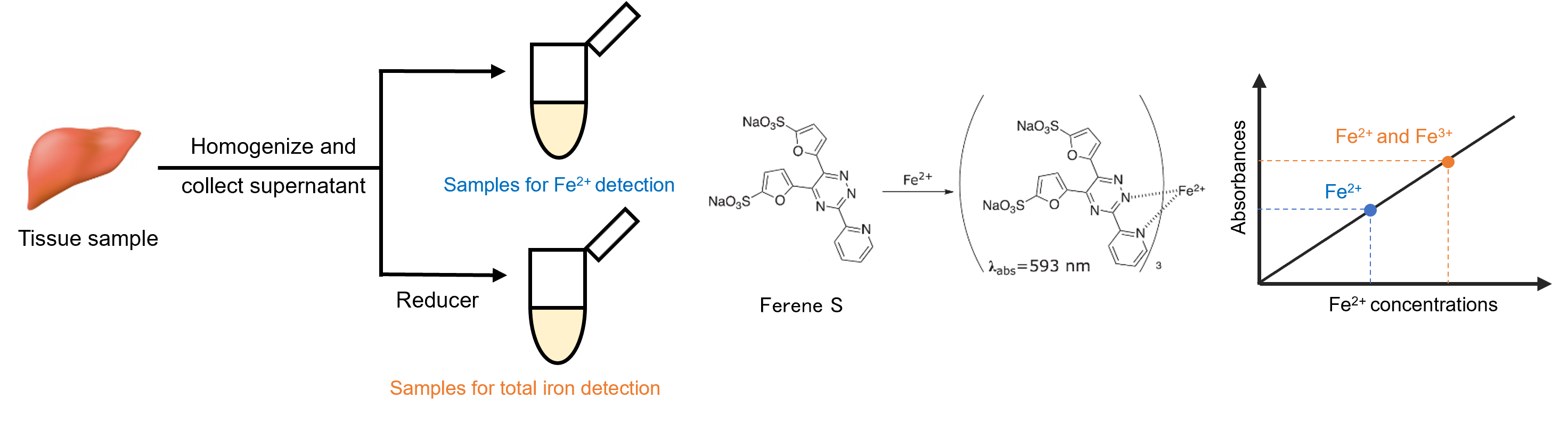
The Iron Assay Kit-Colorimetric is a kit for measuring the amount of iron ions (Fe²⁺ and Fe³⁺) in tissue. It enables data acquisition in approximately 2 h from sample pretreatment to the measurement of iron ion concentration.
The concentration of Fe²⁺ in a tissue sample can be determined by adding Ferene S, which is a chromogenic dye for Fe²⁺, to the tissue lysate and measuring its absorbance. By using the reducing agent included in the kit to reduce Fe³⁺ in the sample to Fe²⁺, the concentrations of total iron and Fe³⁺ in the sample can also be determined.
Ferroptosis Analysis Products
| Product Name | Target | Ditection Properties |
|---|---|---|
| FerroOrange | Intracellular Fe2+ | Microscopy, Plate reader Ex: 543 nm / Em: 580 nm |
| Mito-FerroGreen | Mitochondrial Fe2+ | Microscopy Ex: 505 nm / Em: 535 nm |
| Lyso-FerroRed | Lysosomal Fe2+ | Microscopy, FCM, Plate reader Ex: 551 nm / Em: 571 nm |
| Iron Assay Kit -Colorimetric- | Fe2+ and Fe3+ | Plate reader Colorimetric, λ: 593 nm |
| Liperfluo | Lipid Peroxide | Microscopy, FCM Ex: 488 nm / Em: 500-550 nm |
| Lipid Peroxidation Probe -BDP 581/591 C11- |
Lipid Peroxidation Process |
Microscopy, FCM, Plate reader Pre-reaction, Ex: 488 nm / Em: 510-550 nm Post-reaction, Ex: 561 nm / Em: 600-630 nm |
| MDA Assay Kit | Malondialdehyde | Plate reader Fluorescence, Ex: 540 nm / Em: 590 nm Colorimetric, λ: 532 nm |
| Cystine Uptake Assay Kit | Cystine uptake | Plate reader Ex: 485 nm / Em: 535 nm |
| GSSG/GSH Quantification Kit | GSSG and GSH | Plate reader Colorimetric, λ: 405 nm |
Technical info
Iron Detection Kit and Staining Reagents
Select the appropriate reagent based on your experimental method and detecting equipment.
| Lyso-FerroRed | FerroOrange | Mito-FerroGreen | Iron Assay Kit -Colorimetric- | |
| Intracellular localization | Lysosome | Intracellular | Mitochondria | ー (Tissue) |
|---|---|---|---|---|
| Detection properties | Fluorometric λex : 551 nm, λem : 571 nm |
Fluorometric λex : 543 nm, λem : 580 nm |
Fluorometric λex : 505 nm, λem : 535 nm |
Colorimetric 593 nm |
| Supported devices (filter) |
Microscope, Plate reader, Flow cytometry(TRITC) |
Microscope, Plate reader(Cy3) | Microscope(FITC, GFP) | Plate reader |
| Target | Lysosomal Fe2+ in live cells | Intracellular Fe2+ in live cells | Mitochondrial Fe2+ in live cells | Fe2+ and Fe3+ in tissue |
| Usage count | 35 nmol can be used for 17 assays at 35 mm dish (final concentration 1 µmol/l). |
1 tube (24 µg) can be used for 17 assays at 35 mm dish (final concentration 1 µmol/L) |
1 set (50 µg x 2) can be used for 10 assays at 35 mm dish (final concentration 5 µmol/L) |
50 tests (Make standard curve and detect Fe2+ only: 4 samples, detect Fe2+ and Fe3+: 2 samples) |
Ferrous Ion Selectivity

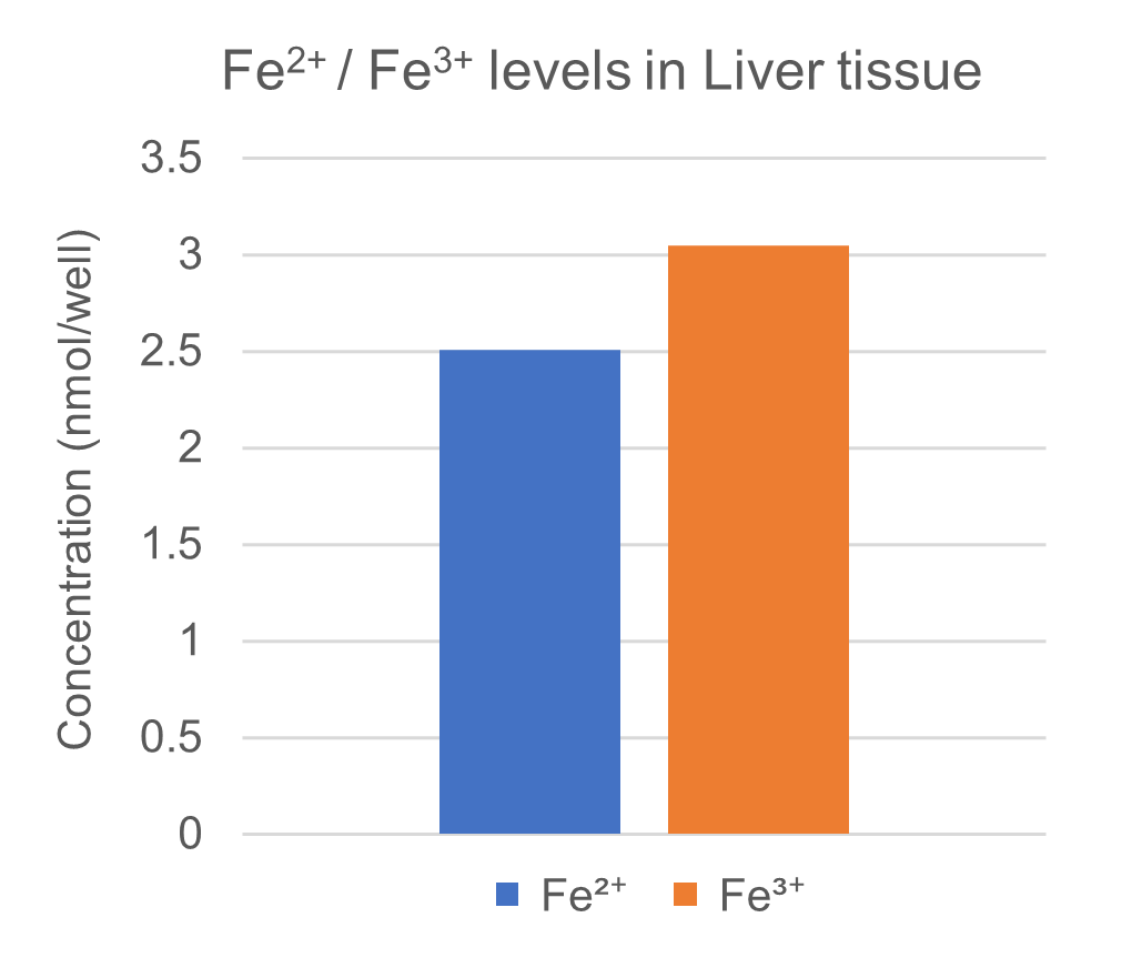
Experimental example: Detection of Fe2+ and Fe3+ in liver sample
Using this kit, we measured the amounts of Fe2+ and Fe3+ in mouse liver. We confirmed that 100 mg of mouse liver contained 2.51 nmol of Fe2+, 3.05 nmol of Fe3+, and a total iron content of 5.56 nmol.