


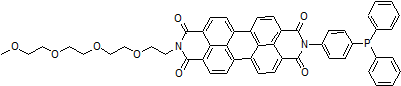
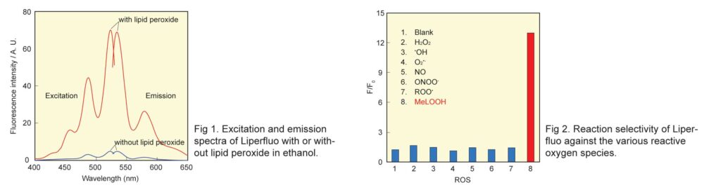
Liperfluo, a perylene derivative containing oligooxyethylene, is designed and exclusively developed by Dojindo for the detection of lipid peroxides. Liperfluo emits intense fluorescence by lipid peroxide specific oxidation in organic solvents such as ethanol. Among fluorescent probes that detect Reactive Oxygen Species(ROS), Liperfluo is the only compound that can specifically detect lipid peroxides. Since the excitation and emission wavelengths of the oxidized Liperfluo are 524 nm and 535 nm, respectively, photo-damage and auto-fluorescence from the sample can be minimized. The tetraethyleneglycol group linked to one end of diisoquinoline ring helps its solubility and dispersibility to aqueous buffer. Liperfluo’s oxidized form is nearly nonfluorescent in an aqueous media and emits a strong fluorescence in lipophilic sites such as in cell membranes. Therefore it can easily be applied to lipid peroxide imaging by a fluorescence microscopy and a flow cytometric analysis for living cells. Liperfluo is used to monitor lipid peroxidation in ferroptosis research.
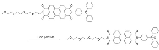
Reaction of Liperfluo with lipid peroxide
Technical info
Live cell imaging of lipid peroxide
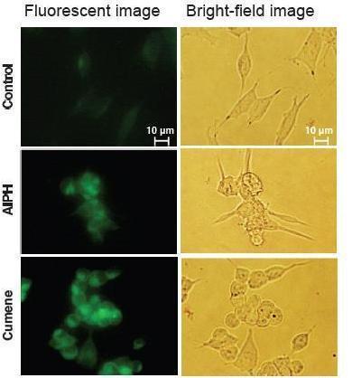
Procedure
1. Innoculate SH-SY5Y cells(6.0 x 10⁵ cells/well) to a 6-well plate.
2. Incubate the plate at 37 ºC for overnight.
3. Add Liperfluo, DMSO solution (final conc. 20 μM) and incubate at
37 ºC for 15 min.
4. Add either Cumene Hydroperoxide (final conc. 100 μM) or AIPH*(final conc. 6 mM).
5. Incubate at 37 ºC for 2 hours.
6. Observe fluorescent by microscope**.
* AIPH: 2,2 Eazobis-[2-(2-imidazolin-2-yl)propane]dihydrochloride
** Olympus IX-71 epifluorescent microscope, mirror unit: U-MNIBA3, exposure time: 10 sec, ISO: 800
Data was kindly provided from Dr. N. Noguchi, Doshisha University, System Life Science Laboratory.
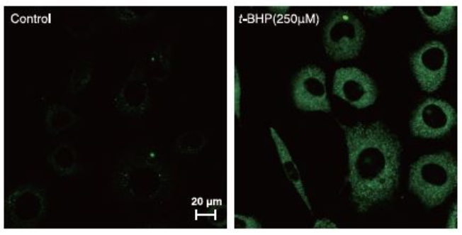
Cell line: L929
Microscope: Zeiss LSM510META
Filter type: FITC(GFP, Alexa488)wide filter
HFT UV/488
NFT490
BP505-550
Procedure
1. Prepare cell suspension (2.5 x 10⁵ cell/well) in 35mm Glass bottom dish and incubate at 37oC overnight in CO2.
2. Discard the media and add new media containing Liperfluo (final conc. 1μM) .
3. Incubate at 37oC for 30 min in CO2.
4. Discard the media add new media containing t-BHP (final conc. 250μM ).
5. Incubate at 37oC for 2 hours in CO2.
6. Observe using confocal microscope.
Data was kindly provided from Dr. T. Kumagai and Dr. H. Imai, Kitasato University, School of Pharmacy.
Flow cytometric analysis of lipid hydroperoxides in live cell
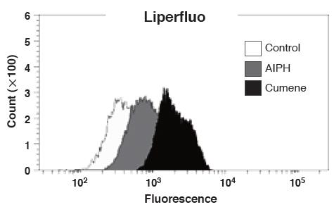
Procedure
1. Innoculate SH-SY5Y cells (6.0 x 10⁵ cells/well) to a 6-well plate.
2. Incubate the plate at 37 ºC for overnight.
3. Add Liperfluo, DMSO solution (final conc. 20 μM) and incubate at 37 ºC for 15 min.
4. Add either Cumene Hydroperoxide (final conc. 100 μM) or AIPH*(final conc. 6 mM).
5. Incubate at 37 ºC for 2 hours.
6. Wash cells with PBS.
7. Collect cells with PBA and analyse by flow cytometer**.* AIPH: 2,2 Eazobis-[2-(2-imidazolin-2-yl)propane]dihydrochloride** BD FACSAriaTM I
Data was kindly provided from Dr. N. Noguchi, Doshisha University, System Life Science Laboratory.
Examples of use in Ferotosis Research
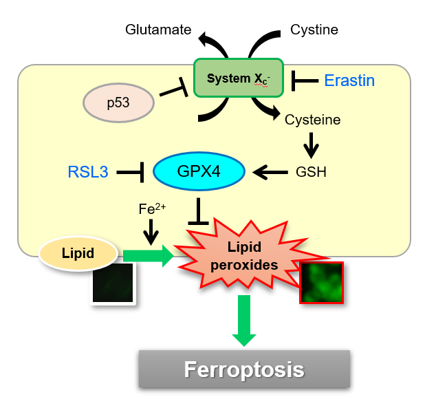
Necrosis, apoptosis and autophagy is known as cell death-related processes. In 2012, Ferroptosis was proposed as one of new cell deaths. Ferroptosis is studied as non apoptotic cell death caused by accumulation of iron ion-dependent lipid peroxide. Liperfluo is used as a fluorescent prove which can detect intracellular lipid peroxide directly.
Ferroptosis: A Regulated Cell Death Nexus Linking Metabolism, Redox Biology, and Disease.
B. R. Stockwell et al., Cell, 2017, 171(2), 273.
Oxidized Arachidonic/Adrenic Phosphatidylethanolamines Navigate Cells to Ferroptosis
V. E. Kagan et al., Nat. Chem. Biol., 2017, 13, (1), 81.
Induction of Ferroptosis by Erastin
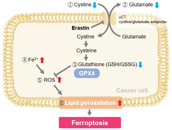
Erastin is a known inducer of ferroptosis. By inhibiting the cystine transporter (xCT), erastin inhibits the uptake of cystine. Cystine is the raw material for GSH. Therefore, Erastin ultimately decreases the amount of GSH. Decreased GSH then results in lipid peroxide accumulation and induction of ferroptosis.
The following experimental examples show changes in each aforementioned index as a consequence of erastin stimulation. Measurements are made using Dojindo reagents.
Using erastin-treated A549 cells, we measured intracellular Fe2+, ROS, lipid peroxide, glutathione, glutamate release into the extracellular space, and cystine uptake. As a result, inhibition of xCT by elastin was observed and also the release of glutamate and uptake of cystine were decreased. Furthermore, elastin treatment decreased intracellular glutathione while it increased intracellular Fe2+ , ROS, and lipid peroxides.

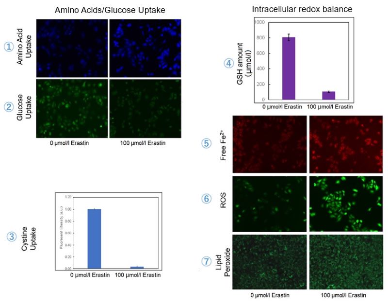
| 1 Amino Acid Uptake | : Amino Acid Uptake Assay Kit (Code: UP04) |
| 2 Glucose Uptake | : Glucose Uptake Assay Kit-Green (Code: UP02) |
| 3 Cystine Uptake | : Cystine Uptake Assay Kit (Code: UP05) |
| 4 Intracellular glutathione | : GSSG/GSH Quantification Kit (Code: G257) |
| 5 Intracellular labile Fe | : FerroOrange (Code: F374) |
| 6 Intracellular total ROS | : ROS Assay Kit -Highly Sensitive DCFH-DA- (Code: R252) |
| 7 Lipid Peroxides | : Liperfluo (Code: L248) |
Cell Line: A549
Incubation Conditions: 100 μmol/l Erastin/MEM, 37℃, 3h