

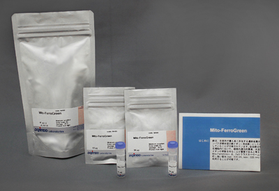
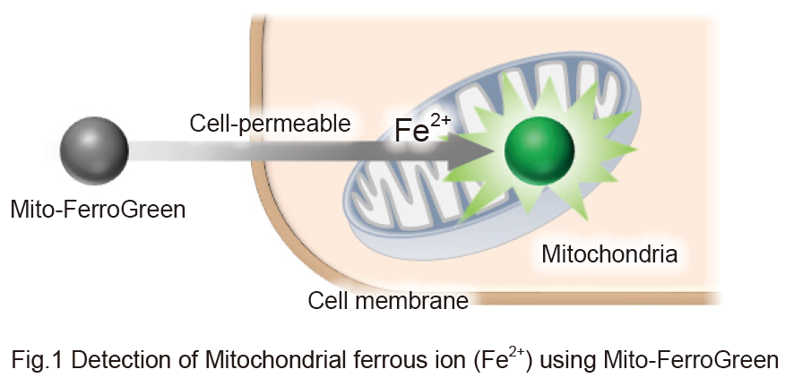
It is reported that iron is the most abundant transition metal element within an organism and shows various physiological activities. Recently, free iron in living cells is getting attention because its high reactivity is suggested to be related to cellular damage or death. Free iron exists in its stable redox states, ferrous ion (Fe2+) and ferric ion (Fe3+). In living cells, it is considered that understanding the behavior of Fe2+ is more important than that of Fe3+ because of the intracellular reductive environment, metal transporters and water solubility of Fe2+. Mito-FerroGreen is a novel fluorescent probe for the detection of ferrous ion (Fe2+) in mitochondria where Fe-S clusters and heme proteins are synthesized, and enables live cell fluorescent imaging of intracellular Fe2+. Mito-FerroGreen has no no chelating ability. Mito-FerroGreen and Fe2+ react irreversibly, which is different from the detection principle of calcium-ion probes such as Fluo 3.
Technical info
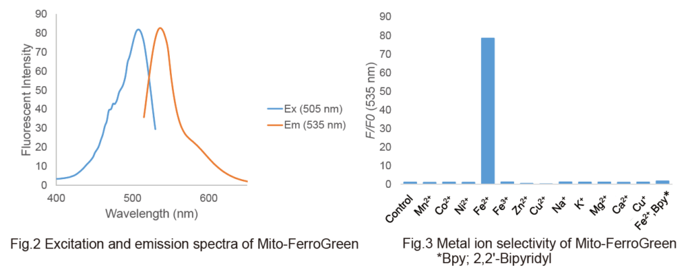
Detection of Mitochondrial ferrous iron (Fe2+) using Mito-FerroGreen
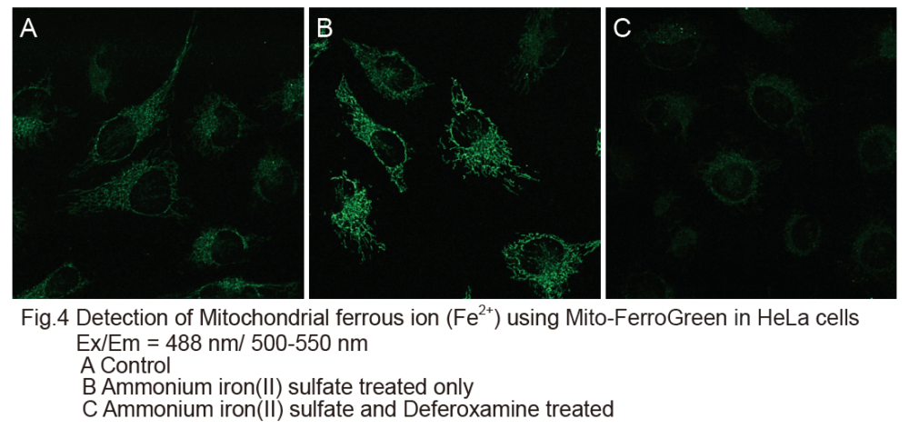
1. HeLa cells were seeded on μ-slide 8 well (ibidi) and cultured at 37oC overnight in a 5% CO2 incubator.
2. The cells were washed with HBSS three times.
3. Mito-FerroGreen working solution (5 μmol/l, 200 μl) were added to the cells, and the cells were incubated at 37oC for 30 minutes in a 5% CO2 incubator.
4. After supernatant was discarded, 10 mmol/l deferoxamine mesylate salt (sigma) prepared with HBSS was added to the cells, and the cells were incubated at 37oC for 30 minutes in a 5% CO2 incubator.
5. The supernatant was discarded and the cells were washed with HBSS three times. After the HBSS was removed, serum-free medium was added to the cells.
6. Ammonium iron(II) sulfate prepared with purified water (10 mmol/l) was added to the cell to be the final concentration of 100 μmol/l.
7. The cells were incubated at 37oC for 1 hour in a 5% CO2 incubator, and the cells were washed with HBSS three times.
8. The cells were observed by confocal fluorescence microscopy.
Double staining with mitochondrial staining probe
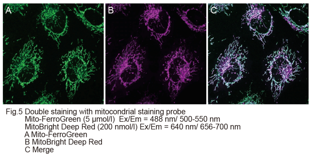
1. HeLa cells were seeded on μ-slide 8 well (ibidi) and cultured at 37oC overnight in a 5% CO2 incubator.
2. The cells were washed with HBSS three times and the supernatant was removed.
3. HBSS (200 μl) containing the final concentration of 5 μmol/l Mito-FerroGreen and 200 nmol/l MitoBright Deep Red (Dojindo※) were added to the cells and the cells were incubated at 37oC for 30 minutes in a 5% CO2 incubator.
4. The cells were washed with HBSS three times.
5. After the HBSS was removed, 200 μl of serum-free medium was added to the cells, and 10 mmol/l ammonium iron(II) sulfate prepared with purified water was added to be the final concentration of 100 μmol/l.
6. The cells were incubated at 37oC for 1 hour in a 5% CO2 incubator, and the cells were washed with HBSS three times.
7. The cells were observed by confocal fluorescence microscopy.
※Our sincere apology that we discontinued distributing MitoBright Deep Red.
Please refer to MitoBright LT Deep Red (Code#:MT12) which improved retention ability.