


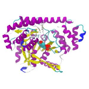
The gene of β-galactosidase from E. coli is widely used as a reporter gene assay marker. Although X-gal is well known reagent to detect β-galactosidase in cell or tissue samples, the assay using these reagents require to fix cells or tissues due to the poor cell-permeability. In addition, so far developed the assay using fluorescence reagents cannot clearly differentiate β-galactosidase-expressed cells or regions.
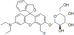
To overcome these issues, Urano, Kamiya and co-workers have successfully developed SPiDER-βGal. SPiDER-βGal ideally possesses cell-permeability and the ability to retain in intracellular region.1)
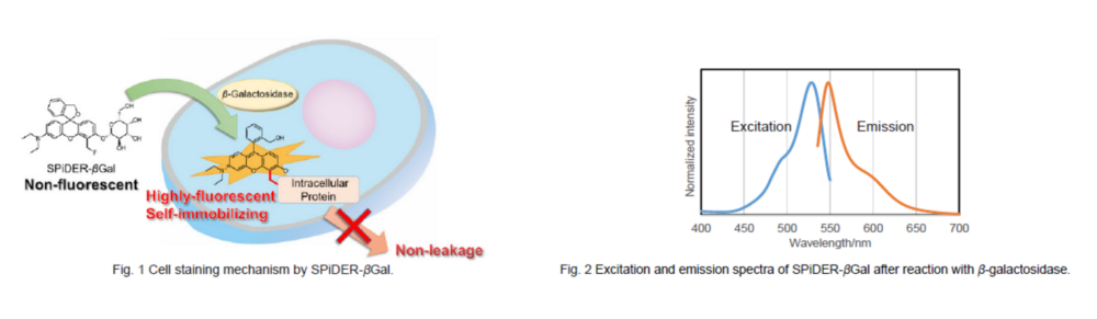
By the enzymatic reaction, SPiDER-βGal immediately forms a quinone methide that acts as electrophile when proteins containing nucleophilic functional groups nearby the molecules. By the probe undergoes the reaction with a protein, the conjugates become fluorescent compounds. Thus, SPiDER-βGal allows a single-cell analysis because it does self-immobilizing to the intracellular proteins.
Cellular Senescence Analysis Products
| Product Name | Detection | Sample | Dyes / Fluorescence Properties |
|---|---|---|---|
| Cellular Senescence Detection Kit – SPiDER-βGal | Microscopy or FCM | Living / Fixed cells | SPiDER-βGal Ex: 500–540 nm / Em: 530-570 nm |
| Cellular Senescence Detection Kit – SPiDER Blue | Microscopy, FCM or Plate reader | Fixed cells | SPiDER Blue Ex: 350-450 nm / Em: 400-500 nm |
| SPiDER-βGal | Microscopy | Tissue | SPiDER-βGal Ex: 500–540 nm / Em: 530-570 nm |
| Cellular Senescence Plate Assay Kit – SPiDER-βGal | Plate reader | Living cells | SPiDER-βGal Ex: 500–540 nm / Em: 530-570 nm |

Technical info
Detection of SA-β-gal in Tissue Samples
A paper on the detection of SA-β-gal in tissue samples from diabetic model mice using SPiDER-βGal has been published.
Quick-frozen tissue was sliced, immersed in 4% paraformaldehyde, and incubated at room temperature for 20 minutes. Then, 20 μmol/L SPiDER-βGal was added to the washed sample in PBS and incubated at 37°C for 1 hour. Finally, the samples were washed with PBS and observed.
For details of the experimental manipulations and data, please refer to this reference.
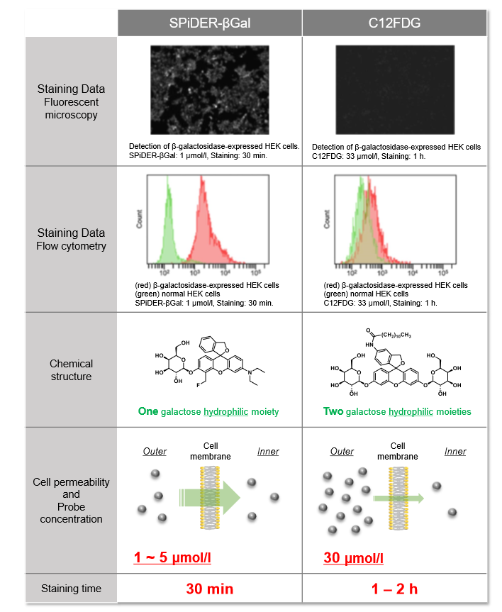
Difference between SPiDER-βGal and C12FDG
Usage Examples:
Fluorescence microscopic detection of β-galactosidase-expressed cells
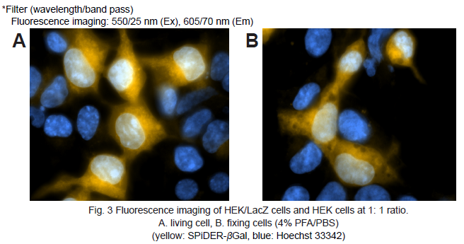
1. HEK cells at 5 × 10⁵ cells/ml (500 μl) and HEK/LacZ cells at 5 × 10⁵ cells/ml (500 μl) were seeded in a 35 mm dish in DMEM (10% fetal bovine serum, 1% penicillin-streptmycin) and cultured overnight in a 5% CO2 incubator at 37oC.
2. The cells were washed with 2 ml of Hanks’ HEPES buffer twice.
3. SPiDER-βGal working solution (2 ml) was added to the culture dish. The cells were then incubated for 15 minutes at 37oC.
4. After the supernatant was removed, the cells were washed Hanks’ HEPES buffer (2 ml) twice.
5. Hanks’ HEPES buffer (2 ml) were added and the cells observed under a fluorescence microscope. (Fig. 3A)
6. After the supernatant was removed, 4% paraformaldehyde (PFA) /PBS solution (2 ml) was added to the culture dish. The cells were then incubated for 15 minutes at room temperature.
7. After 4% PFA/PBS solution was removed, the cells were washed Hanks’ HEPES buffer (2 ml) twice.
8. Hanks’ HEPES buffer (2 ml) were added and the cells observed under a fluorescence microscope. (Fig. 3B)
Flow cytometric detection of β-galactosidase-expressed cells
1. HEK cells at 5 × 10⁵ cells/ml (500 μl) and HEK/LacZ cells at 5 × 10⁵ cells/ml (500 μl) were mixed in a microtube.
2. SPiDER-βGal DMSO stock solution (1 μl) was added to the tube. The cells were then incubated 15 minutes at 37oC .
3. The cells were analyzed under a flow cytometer. (488 nm excitation, 530/30 nm bandpass filter)
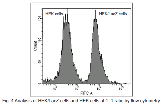
β-galactosidase-expressed cells (HEK/LacZ cells) were clearly differentiate from HEK cells in flow cytometry data analysis.
Live Imaging of Drosophila tissue
Living drosophila tissue was incubated with 10 µmol/l SPiDER-βGal and 16 µmol/l Hoechst 33342 for 20-30 min, observed with confocal microscope.
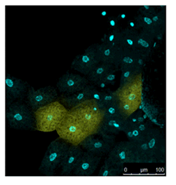
Data was kindly provided by Dr. Y. Urano, at University of Tokyo, Graduate School of Medicine.
Imaging of a Fixed Tissue of a Drosophila
Formalin fixed drosophila generative cells were stained by SPiDER-βGal and DAPI. The β-galactosidase, which is expressed at the nucleus of drosophila generative cells, could be obtained by SPiDER-βGal.
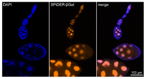
Data was kindly provided by Dr. T. Nakamura, at University of Kumamoto, Institute of Molecular Embryology and Genetics.
Imaging of a Pancreas of a Mouse
SPiDER-βGal and Salmon-Gal were evaluated in the detectability of β-galactosidase on a pancreas of a mouse. SPiDER-βGal was able to detect the difference of the expression point of β-galactosidase more clearly than Salmon-Gal.
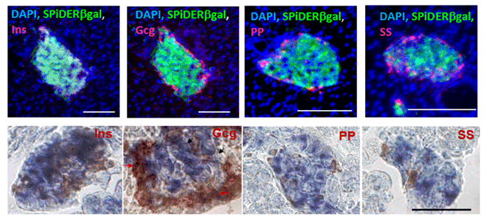
Data was kindly provided by Dr. S. Kume, at Tokyo Institute of Technology, School of Bioscience and Biotechnology.