




Amino acids are essential for intracellular protein and nucleic acid synthesis, Especially for cancer cells which are proliferating continually. Since the supply of acetyl CoA from the glycolytic pathway is decreased in cancer cells, they have a furthermore huge demand for amino acids which are an important nutrient source for the TCA cycle. The research has shown that cancer cells increase the expression of the amino acid transporter LAT1 (L-type amino acid transporter 1) and take up a large number of amino acids, which is expected to be a new target for anti-Cancer drugs’ discovery.
In immunotherapy, not only the metabolic changes in cancer cells but also in immune cells are considered to affect the treatment. For example, as senescence occurs in immune cells, their metabolic system changes, and their immune capacity to attack cancer cells decreases. Therefore, research is being conducted on nutrient uptake regulation by immune cells and improving the effectiveness of cancer immunotherapy.
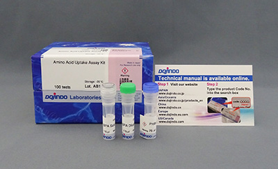
After amino acid analogs (BPA) are taken up into cells via amino acid transporters, the fluorescent probe permeates the cell membrane and binds to the amino acid analogs, emitting fluorescence (λex=360 nm, λem=460 nm). This product is suitable for fluorescence imaging, plate reader, and flow cytometry, making it possible to visualize and quantify the amino acid uptake capacity of cells and is useful for evaluation of amino acids uptake capacity and screening of amino acids transporter inhibitors.
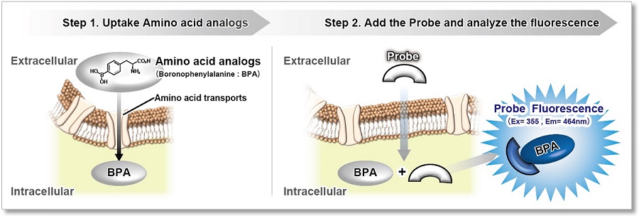

Technical info
Evaluate the inhibitor of amino acids transporter and compare the amino acids uptake capacity between different cell lines. 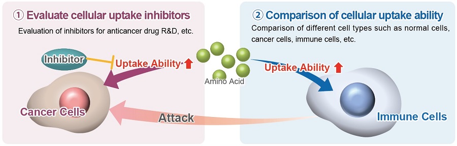
Comparison with Existing Method
Compare to the existing method, the required time can be greatly reduced.
| Method | Instrument | Time | Remarks |
|---|---|---|---|
| Amino Acid Uptake Assay Kit | Fluorescent Microscope Plate Reader Flow Cytometer |
60 min | Simple procedure for amino acid analogs uptaking analysis |
| Radioisotope Labeling | Liquid Scintillation | 2 Days | Allow testing different types of amino acids, but there are restrictions on use. |
| Metabolomics | LC/MS | 3 hr | Allow testing different types of amino acids at the same time, but the result is hard to analyze. |
Application Data: Evaluation of BCH (Inhibitor of amino acids transporter)
Evaluate the inhibition ability BCH (2-Aminobicyclo[2.2.1]heptane-2-carboxylic acid) in HeLa cells with Amino Acid Uptake Assay Kit.
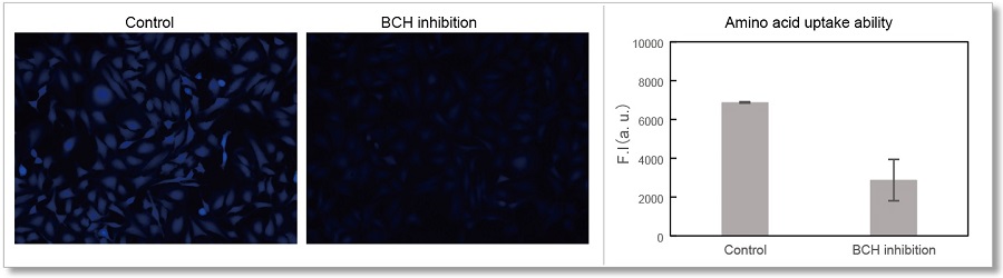
<Experimental Conditions>
Cell Line: HeLa cells
Medium: MEM (5.5 mmol/l Glucose)
Incubation: 1 mmol/l BCH/HBSS (Hanks’ Balanced Salt Solution), 37℃, 30 min
Instrument:Fluorescent Microscopy(Ex=340-380 nm, Em: 435-485 nm)
Instrument:Plate Reader (Ex=360 nm, Em: 460 nm)

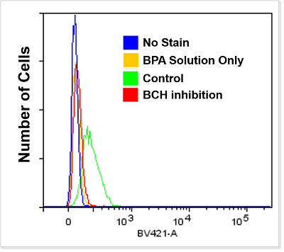
<Experimental Conditions>
Instrument: Flow Cytometer (Ex=405 nm, Em: 425-475 nm)
Branched Chain Amino Acids (BCAA) and Disease
It is clear that the intracellular metabolism of branched-chain amino acids (BCAA) is involved in various diseases such as cancer, aging, diabetes, and obesity. BCAA is mainly taken up into cells via neutral amino acid transporter (LAT).
The amino acid analog (BPA) used in this kit has been confirmed to be taken up via LAT in the same way as BCAA, and this kit can be used to evaluate the intracellular uptake of BCAA.
Cancer cells
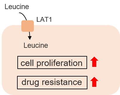
Recent studies have shown that breast cancer cells take up large amounts of leucine, a BCAA, via LAT1. the BCAA uptake transporter LAT1 is expected to be a target for new breast cancer drugs.
Saito, Y., Li, L., Coyaud, E., et al, “LLGL2 rescues nutrient stress by promoting leucine uptake in ER+ breast cancer.” Nature, 2019, 569, 275.
Cellular senescence
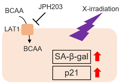
Cells are known to undergo cellular senescence upon X-irradiation via DNA damage and ROS generation. The combination of X-irradiation and LAT1 inhibitors in cancer cells has been shown to promote cellular senescence. The importance of BCAAs in cellular senescence has been suggested.
Tomoki Bo et al.,” LAT1 inhibitor JPH203 sensitizes cancer cells to radiation by enhancing radiation-induced cellular senescence”, Translational Oncology, 2021, 14, 101212.
Obesity
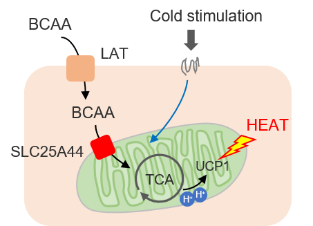
Brown adipose tissue (BAT) during cold exposure takes up BCAAs for heat production. It has been shown that intracellularly taken-up BCAAs are oxidized in mitochondria and contribute to the heat production of BAT.
Yoneshiro T, et al., “BCAA catabolism in brown fat controls energy homeostasis through SLC25A44”, Nature, 2019, 572, 614.
Experimental Example: Evaluation with BCAA (Leucine)
The addition of L-Leucine, a BCAA, and BCH, a LAT1 inhibitor, inhibited the uptake of BPA into the cells.
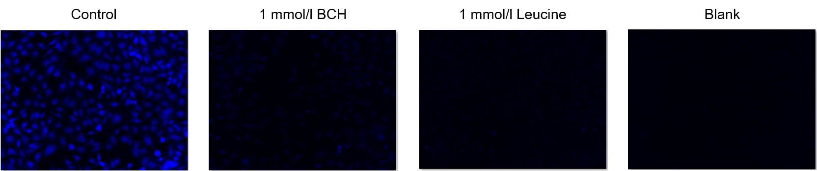
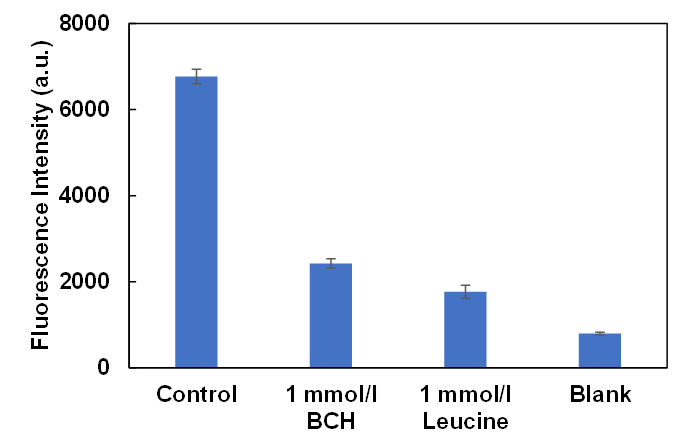
Cells: HeLa
Inhibitors: LAT1 inhibitor BCH (2-Aminobicyclo[2.2.1]heptane-2-carboxylic acid) and L-Leucine
Experimental Example: Evaluation of Amino Acid Transporter Overexpressing Cells
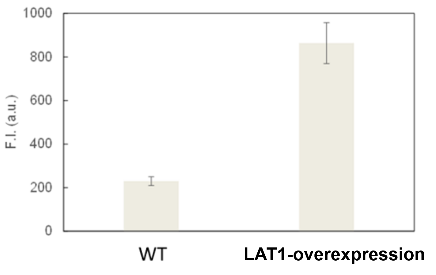
Amino acid uptake in LAT-1-overexpressing MCF7 cells was greatly increased compared with the wild type.
Courtesy of Dr. Yasuhiro Saito (Institute for Advanced Biosciences, Keio University)
Experimental Example: Evaluation by Amino Acid Transporter Inhibitor (T3)
BPA cellular uptake was inhibited in a concentration-dependent manner by the addition of T3, an LAT1 inhibitor.
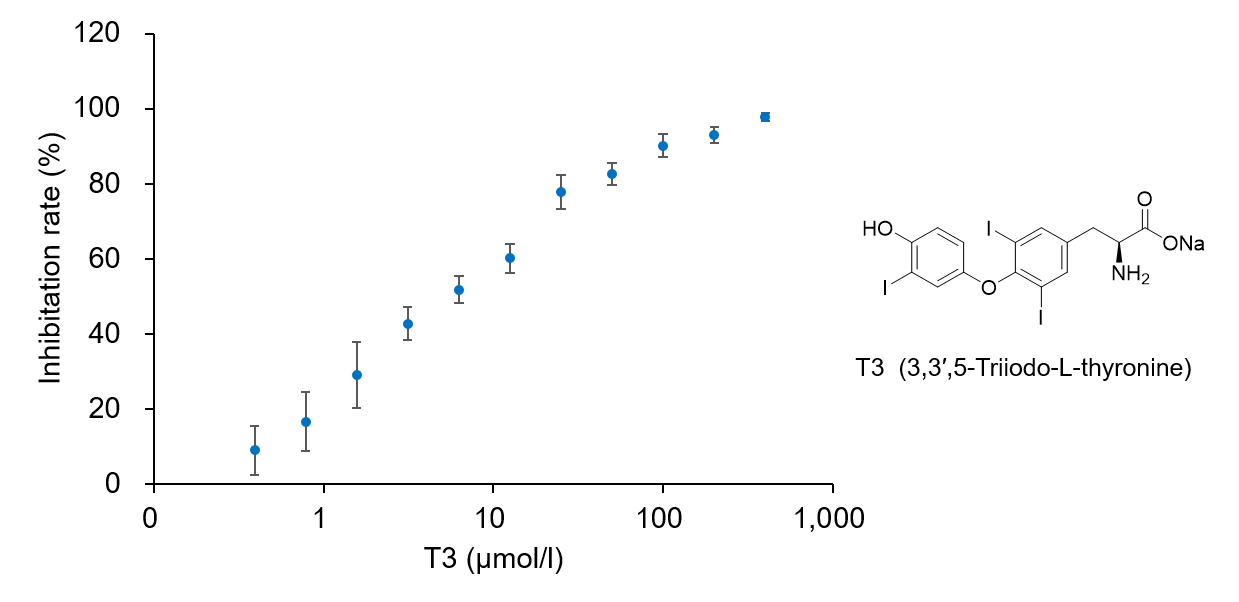
Cells: HeLa
Inhibitors: LAT1 inhibitor T3 (3,3′,5-Triiodo-L-thyronine)
Experimental Examole: Induction of Ferroptosis by Erastin
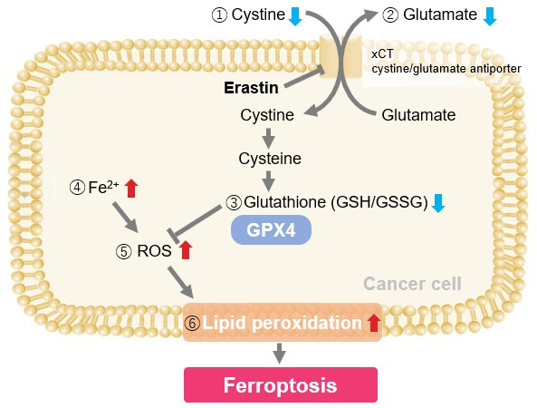
Erastin is a known inducer of ferroptosis. By inhibiting the cystine transporter (xCT), erastin inhibits the uptake of cystine. Cystine is the raw material for GSH. Therefore, Erastin ultimately decreases the amount of GSH. Decreased GSH then results in lipid peroxide accumulation and induction of ferroptosis.
The following experimental examples show changes in each aforementioned index as a consequence of erastin stimulation. Measurements are made using Dojindo reagents.
Using erastin-treated A549 cells, we measured intracellular Fe2+, ROS, lipid peroxide, glutathione, glutamate release into the extracellular space, and cystine uptake. As a result, inhibition of xCT by elastin was observed and also the release of glutamate and uptake of cystine were decreased. Furthermore, elastin treatment decreased intracellular glutathione while it increased intracellular Fe2+ , ROS, and lipid peroxides.

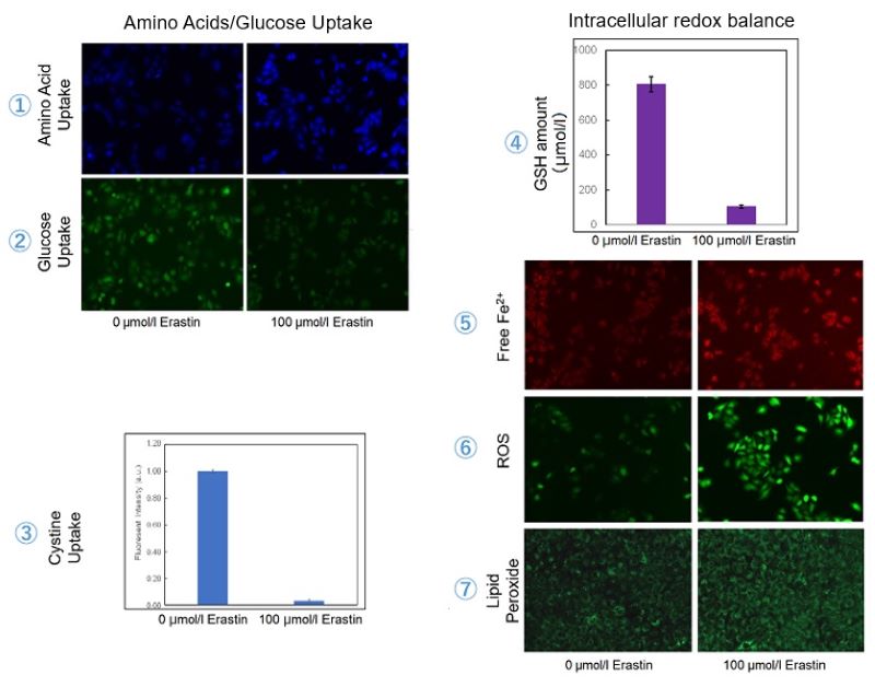
| 1 Amino Acid Uptake | : Amino Acid Uptake Assay Kit (Code: UP04) |
| 2 Glucose Uptake | : Glucose Uptake Assay Kit-Green (Code: UP02) |
| 3 Cystine Uptake | : Cystine Uptake Assay Kit (Code: UP05) |
| 4 Intracellular glutathione | : GSSG/GSH Quantification Kit (Code: G257) |
| 5 Intracellular labile Fe | : FerroOrange (Code: F374) |
| 6 Intracellular total ROS | : ROS Assay Kit -Highly Sensitive DCFH-DA- (Code: R252) |
| 7 Lipid Peroxides | : Liperfluo (Code: L248) |
Cell Line: A549
Incubation Conditions: 100 μmol/l Erastin/MEM, 37℃, 3h