








Adenosine triphosphate (ATP) is an energy source for living cells and is synthesized by both glycolysis and mitochondrial oxidative phosphorylation. Mitochondria generate 95% of cellular ATP, and mitochondrial dysfunction reduces ATP levels in cells. Therefore, the measurement of ATP levels has been established as a proxy for mitochondrial activity. Decreased ATP levels are associated with cancer, aging, neurodegenerative diseases, and mitochondrial diseases. Cancer cells rely on glycolysis, a process that is less efficient than oxidative phosphorylation to synthesize ATP. However, recent studies have revealed that ATP synthesis in cancer cells shifts from glycolysis to oxidative phosphorylation when glycolysis is suppressed.
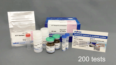
The ATP Assay Kit-Luminescence enables the quantitation of intracellular ATP by luciferase luminescence assay. This kit can be applied for microplate assay. Furthermore, this kit can be applied for microplate assay.

Technical info
The ATP Assay Kit-Luminescence enables the quantitation of intracellular ATP by luciferase luminescence assay. This kit can be applied for microplate assay. Furthermore, this kit can be applied for microplate assay.
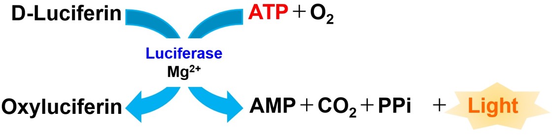
Generate a stable luminescent signal with high reproducibility
Without complicated steps such as medium removal and cell washing, the ATP Assay Kit-Luminescence can generate a stable luminescent signal with a half-life of greater than 3 hours by simply mixing the kit components and adding the reagent. This kit contains an ATP Standard, which provides highly reproducible data without measuring unstable ATP.
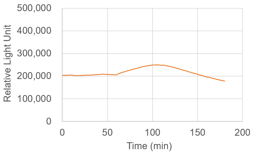
Time-course measurements of relative light units collected 3 hours after the addition of the reagent.
Cell type:HeLa cells (10,000cells/well)
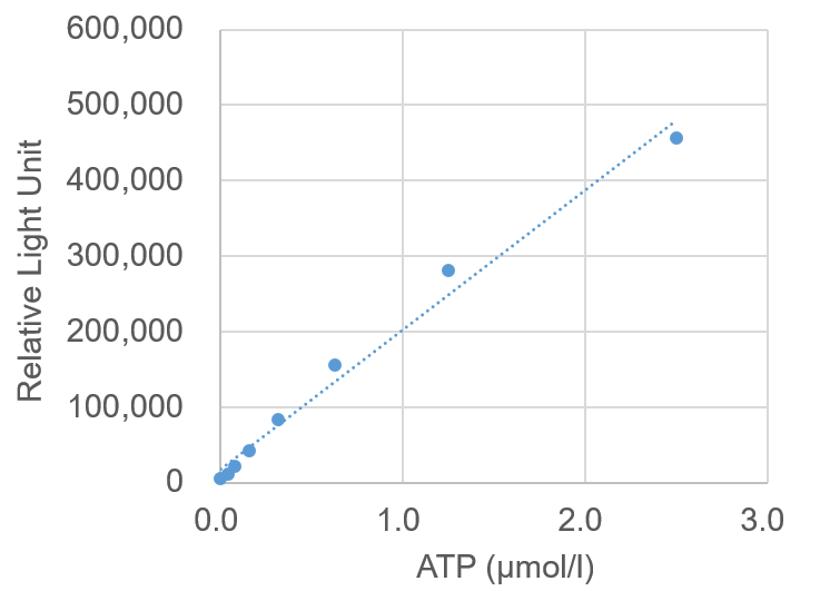
Preparation of Calibration Curve
ATP levels in a sample can be measured by a calibration curve established with ATP Standards included in this kit. If the ATP levels are greater than or equal to 2.5 µmol/l, the sample must be diluted before measurement.
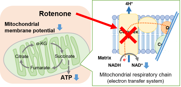
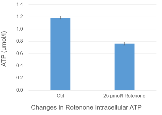
Change in intracellular metabolism of rotenone-treated cells
Rotenone, which is known to inhibit the mitochondrial electron transport chain, was added to Jurkat cells, followed by measurement of intracellular ATP using the ATP Assay Kit-Luminescence. As a result, ATP production in the mitochondrial respiratory chain (the electron transport chain) was inhibited, and ATP concentrations were lower than those in the control cells.
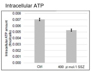
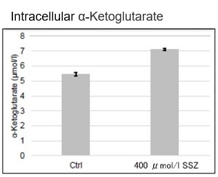
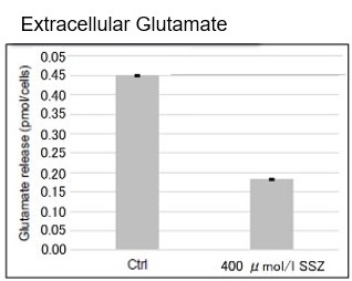
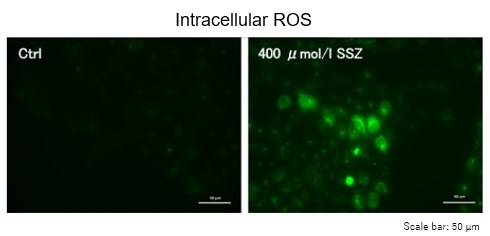
Sulfasalazine Alters Intracellular Metabolites and Increases ROS in A549 Cells
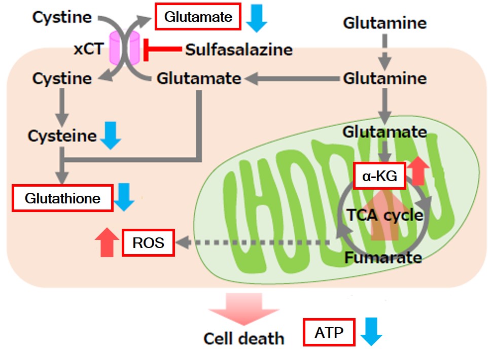 |
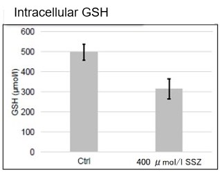
After addition of sulfasalazine (SSZ), a known inhibitor of cystine/glutamate transporter (xCT), to A549 cells, we observed changes in intracellular glutathione (GSH), ATP, α-ketoglutarate (α-KG), ROS and glutamate release. The results indicated that the addition of SSZ The results showed that the addition of SSZ decreased intracellular ATP, glutathione (GSH) and glutamate release, and increased intracellular α-ketoglutarate and ROS. Cells: A549 cells (1 x 10⁶ cells) Exposure time: 48 hours Product in Use
Reference |
     |
|
Experimental Example: Change in Metabolism in Liver Tissue of MASH-Induced Mouse
It is known that metabolic dysfunction-associated steatohepatitis (MASH) results in decreased ATP, α-ketoglutarate (α-KG), and NAD levels in affected tissues. ATP, α-KG, and NAD levels were measured in the liver tissue of type 1 diabetic model mice (STAM model) that were treated with a high-fat diet (MASH induction) since 4 weeks old. Measurement at 10 weeks old confirmed that ATP, α-KG, and NAD levels indeed decreased in the tissue samples after MASH induction.
 Note: for more details about the experimental procedure, please refer to Q&A “Are there any examples of experiments using tissue samples?”
・Cellular ATP :ATP Assay Kit-Luminescence (Code:A550).
・Cellular α-KG:α-Ketoglutarate Assay Kit-Fluorometric (Code:K261).
・Cellular NAD :NAD/NADH Assay Kit-WST (Code:N509).
References:
| ATP | Francesco Bellanti, et al., “Synergistic interaction of fatty acids and oxysterols impairs mitochondrial function and limits liver adaptation during nafld progression”, Redox Biology, 2018, 15, 86-96. |
|---|---|
| α-KG | Jianjian Zhao, et al., “The mechanism and role of intracellular α-ketoglutarate reduction in hepatic stellate cell activation”, Bioscience Reports, 2020, 40, (3). |
| Ali Canbay, et al., “L‑Ornithine L‑Aspartate (LOLA) as a Novel Approach for Therapy of Non‑alcoholic Fatty Liver Disease”, Drugs, 2019, 79, 39-44. | |
| NAD | Jinhan He, et al., “Activation of the Aryl Hydrocarbon Receptor Sensitizes Mice to Nonalcoholic Steatohepatitis by Deactivating Mitochondrial Sirtuin Deacetylase Sirt3”, Mol. and Cell. Biol., 2013, 33, (10), 2047-55. |