

Cytotoxicity Assay
- Can be used with and without transferring supernatant
- Stable Working Solution (6 months at 0-5 oc)
- No need to freeze the Working Solution
Detection Principle
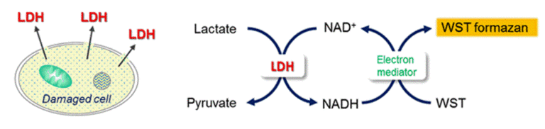
Lactate dehydrogenase(LDH) is an enzyme that presents in almost cell types and it catalyzes the oxidation of lactate to pyruvate in the presence of co-enzyme NAD+. Once cells are impaired by stress, injuries, chemicals, or intercellular signals, LDH is rapidly released from the cell membrane. Thus, the measurement of the amount of released LDH from cells is one of the major methods to assess the cell death. Since Dojindo’s Cytotoxicity LDH Assay Kit-WST neither reflects the activity of living cells nor is harmful to cells, it allows the assay to perform in wells containing both viable and damaged cells.
| Dojindo Molecular Technologies, Inc.
|
Technical info
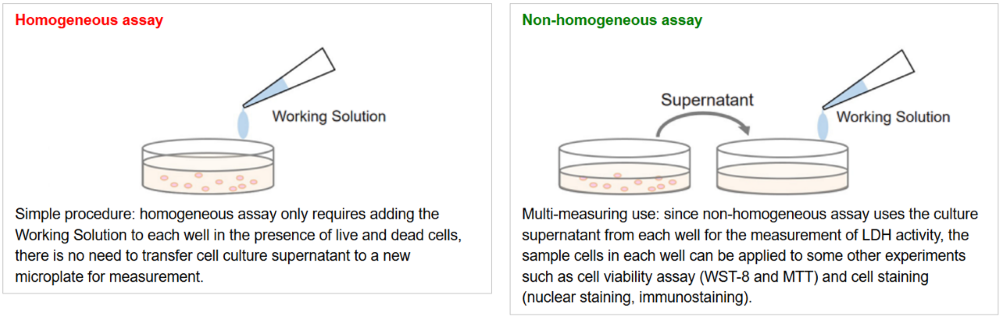
Flexible Application Methods
Cytotoxicity LDH Assay Kit-WST can be used with or without transferring the supernatant. Choose the method that best fits your experiment.
In assays with suspension cells, the method without supernatant transfer eliminates the need for a microplate centrifuge to settle cells at the bottom of the plate.
Video Guide
How to Use the Homogeneous Assay(without transfer) and Non-homogeneous Assay (with transfer)
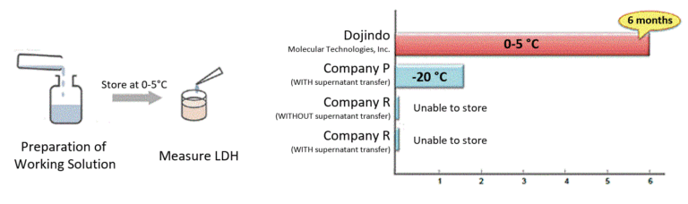
Stable Working Solution
Working Solution is stable for 6 months under refrigerated conditions. Therefore, after the preparation, Working Solution can be used as a ready-to-use solution at any time during this period.
Nearly 90% lower total cost* compared with short-stability LDH kits.
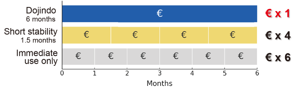
*Based on a comparison of 2000 tests over a continuous six month usage period. Actual results may vary depending on assay frequency and local pricing.
Cytotoxicity of Mitomycin C using HeLa cells
The cytotoxicity of Mitomycin C on HeLa cells was assessed using the Cell Counting Kit-8 and the Cytotoxicity LDH Assay Kit-WST. The Cell Counting Kit-8 reflects intracellular metabolic activity, while the Cytotoxicity LDH Assay Kit-WST detects LDH released from damaged cell membranes. Because the assays measure different indicators, the Mitomycin C concentration at which cytotoxicity was observed varied between the two methods.
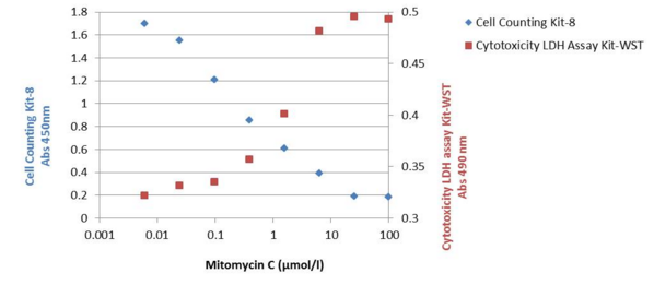
Cell Line: HeLa
Medium: MEM + 10% FBS
Test Compound: Mitomycin C
Exposure: 37 °C, 5% CO₂, 48 h
Detection: Cell Counting Kit-8 (450 nm), Cytotoxicity LDH Assay Kit-WST (490 nm)
Complement-Dependent Cytotoxicity Assay Using RAJI Cells
Complement-dependent cytotoxicity (CDC) was evaluated using RAJI cells. An increase in absorbance was observed in a concentration-dependent manner with the anti-CD20 antibody, indicating that binding of the antibody to the cells activated the complement system and resulted in cell lysis.
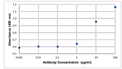
For reference, see the ADCC Assay manual (a related assay method).
Cell Death Related Products
| Type | Product Name | Target | Detection Properties |
|---|---|---|---|
| Cell viability | Cell Counting Kit-8 | Dehydrogenase Activity |
Plate reader Colorimetric, λ= 450nm |
| Necrosis (Cytotoxicity) |
Cytotoxicity LDH Assay Kit-WST | Leaked LDH | Plate reader Colorimetric, λ= 490nm |
| Apoptosis | Annexin V Apoptosis Plate Assay Kit | Phosphatidylserine | Plate reader Ex: 488 nm / Em: 525 nm |
| Ferroptosis | FerroOrange | Intracellular Fe2+ | Microscopy, FCM, Plate reader Ex: 543 nm / Em: 580 nm |
| Ferroptosis | Mito-FerroGreen | Mitochondrial Fe2+ | Microscopy Ex: 505 nm / Em: 535 nm |
| Ferroptosis | Liperfluo | Lipid Peroxide | Microscopy, FCM Ex: 488 nm / Em: 500-550 nm |