


The lysosome is an organelle in which a biomembrane forms an acid vacuole. Lysosomes contain various degrading enzymes and contribute to maintaining intracellular homeostasis by acting as a waste disposal system. Recent findings reveal that lysosomal dysfunction is related to some neurodegenerative disorders. Consequently, the investigation of lysosomal function is attracting considerable interest in the scientific community.
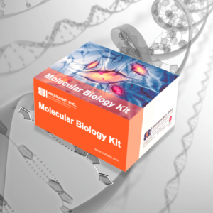
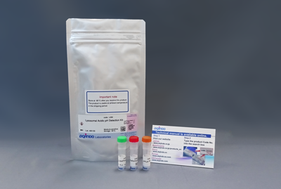
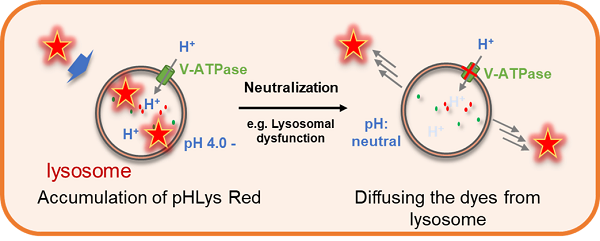
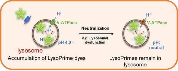
This kit includes lysosome staining dyes, pHLys Red (pH-sensitive), and LysoPrime Green (pH-resistant). The pHLys Red and LysoPrime Green accumulate in the intact lysosomes. The fluorescence intensity of pHLys Red is enhanced as the acidity increases, and weak fluorescence is observed when lysosomes are neutralized due to the lysosomal dysfunction. On the other hand, LysoPrime Green gives stable emissions even lysosomes are neutralized. Lysosomal pH and lysosomal mass can be measured by combining these two dyes.
Lysosomal Analysis Products
| Product Name | Lysosomal pH Detection Dyes and Fluorescence Properties |
Lysosomal Quantity Detection Dyes and Fluorescence Properties |
|---|---|---|
| Lysosomal Acidic pH Detection Kit | pHLys Red Ex: 561 nm / Em: 560-650 nm |
LysoPrime Green Ex: 488 nm / Em: 500-600 nm |
| Lysosomal Acidic pH Detection Kit – Green/Deep Red | pHLys Green Ex: 488 nm / Em: 490-550 nm |
LysoPrime Deep Red Ex: 633 nm / Em: 640-700 nm |
| pHLys Red – Lysosomal Acidic pH Detection | pHLys Red Ex: 561 nm / Em: 560-650 nm |
|
| LysoPrime Deep Red – High Specificity and pH Resistance | LysoPrime Deep Red Ex: 633 nm / Em: 640-700 nm |
|
| LysoPrime Green- High Specificity and pH Resistance | LysoPrime Green Ex: 488 nm / Em: 500-600 nm |

Technical info
Comparative Performance Analysis
All data were generated in-house under identical imaging and treatment conditions.
Lysosome tracking dye
| Dye | LysoPrime Green | LysoPrime Deep Red | Lysosomal Tracker Green Major commercial supplier |
|
pH Stability |
Resistant to pH elevation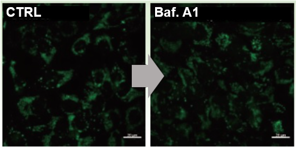 |
Resistant to pH elevation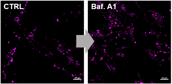 |
Reduced under pH elevation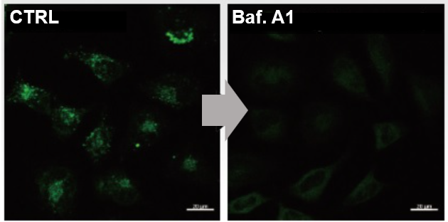 |
|
Lysosomal Retention Time |
Long-term Retention Stable Localization (>24 h) 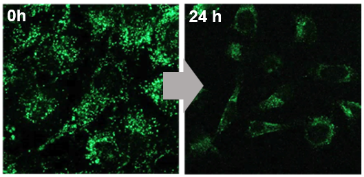 |
Long-term Retention Stable Localization (>24 h) 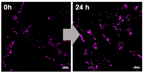 |
Rapid Signal Decline <90 min (identical conditions) 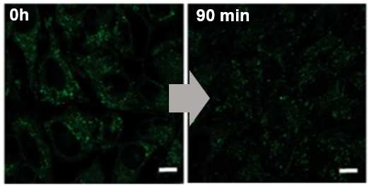 |
Lysosomal pH Sensors
| Dye | pHLys Green | pHLys Red | Lysosomal pH Sensor Green Major commercial supplier |
|
pH Sensitivity |
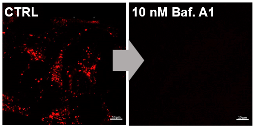
High pH Responsiveness Signal clearly reduced under Baf. treatment 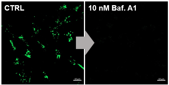 |
High pH Responsiveness Signal clearly reduced under Baf. treatment
|
Less sensitive Incomplete signal suppression under Baf. treatment 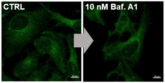 |
|
Lysosomal Retention Time |
Long-term Retention Stable Localization (>24 h) 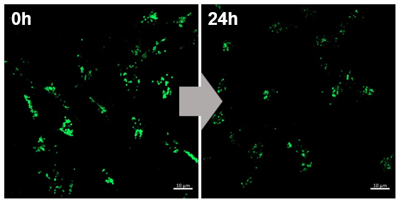 |
Long-term Retention Stable Localization (>24 h) 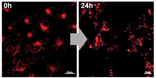 |
Rapid signal decrease <90 min (identical conditions) 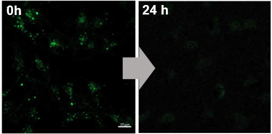
|
Accurate Lysosomal Function Analysis with Two Different Lysosome Fluorescent Probe
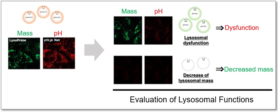
Lysosomes are acidic vesicles surrounded by biological membranes and contain a variety of degradative enzymes. The role of lysosomes is to break down unwanted substances, and they contribute to the maintenance of homeostasis in the body. Since lysosomal dysfunction is deeply involved in the development and progression of neurodegenerative diseases, a detailed analysis of lysosomes is extremely important for elucidating pathological conditions and developing therapeutic drugs. When competitor’s dyes are used to analyze lysosomal function, it is difficult to determine whether the lysosomal mass or their function (pH) has changed because the analysis is based only on the fluorescence intensity of a single dye.
This kit contains two types of dyes: pHLys Red, which shows a lysosomal pH-dependent change in fluorescence intensity, and LysoPrime Green, which is lysosomal pH-resistant. By combining these two dyes, the lysosomal function can be analyzed in detail by simultaneously analyzing lysosomal mass and pH. In addition, the lysosomal acidification inhibitor Bafilomycin A1 is included in this kit, making it easy to obtain negative controls even for first-time researchers.
Outline of Lysosomal function detection with this kit.
pHLys Red: Lysosomal pH-sensitive Fluorescent Probe

LysoPrime Green: Lysosomal pH-resistant Fluorescent Probe

Lysosomal Acidification Inhibitor Included
Fluorescence imaging of lysosomal mass and pH of Bafilomycin A1 (Baf. A1)-induced HeLa cells was performed. The results showed that lysosomal pH was increased in Baf. A1-treated cells compared to control cells, while lysosomal mass was unchanged. Thus, the lysosomal mass and pH was detected at the same time.

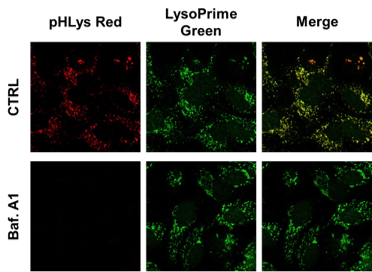
<Experimental Conditions>
LysoPrime Green: Ex = 488 nm, Em = 490 – 550 nm
pHLys Red: Ex = 561 nm, Em = 560 – 620 nm
Application Data: Simultaneously evaluation of lysosomal function and mitochondrial superoxide
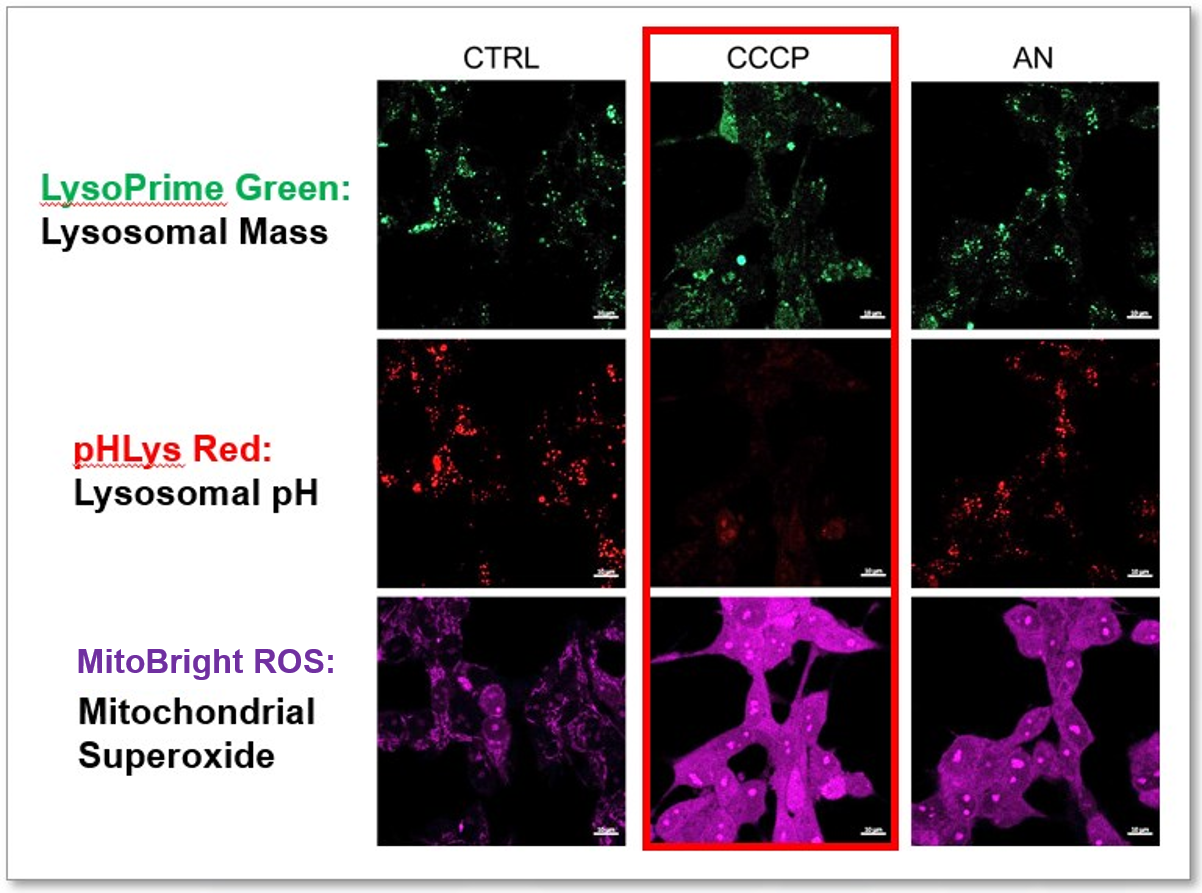
CCCP and Antimycin are recognized inducers of mitochondrial ROS, linked to the loss of mitochondrial membrane potential. Recent studies have shown that CCCP induces not only mitochondrial ROS but also lysosomal dysfunction. To observe mitochondrial ROS, HeLa cells were labeled with MitoBright ROS Deep Red for Mitochondrial Superoxide Detection, and the lysosomal mass and pH were independently detected with LysoPrime Green and pHLys Red. Co-staining with MitoBright ROS and Lysosomal dyes revealed that CCCP, unlike Antimycin, triggers concurrent lysosomal neutralization and mitochondrial ROS induction.
Reference: Benjamin S Padman, et. al., Autophagy (2013)
Products in Use
– LysoPrime Green
– pHLys Red
– Lysosomal Acidic pH Detection Kit
– MitoBright ROS – Mitochondrial Superoxide Detection