









Mitochondria is an important organelle that uses oxygen to synthesize ATP, producing the necessary energy for living cells to thrive1). Decreased mitochondrial activity and mitochondrial dysfunction are associated with cancer, aging, and neurodegenerative diseases such as Alzheimer’s and Parkinson’s disease2,3). Therefore, mitochondrial membrane potential (MMP) has been widely studied as a promising target for mitochondria-related diseases.
Reference
1) K. F. Ferri, et al., J. Exp. Med., 2000, 192, 1081.
2) N. Matsuda, et al., J. Cell Biol., 2010, 189, 211.
3) J. L. Wang, et al., PNAS, 2000, 97, 7124.

Technical info
Comparison of reagents for mitochondrial membrane potential detection
| Features | Sensitivity | Fixation | Monitoring | Fluorescence change (upon loss of mitochondrial membrane potential) | Detection (ex/em) |
|
|---|---|---|---|---|---|---|
| JC-1 (JC-1 MitoMP Detection Kit) |
Recomended for starting-up | ✓ | Color change from red to green | Green: 450-490 nm / 500-550 nm Red: 530-560 nm / 570-640 nm
|
||
| MT-1 (MT-1 MitoMP Detection Kit) |
Recommended for more detailed analysis | ✓ (High) |
✓ | ✓ | Decrease in fluorescence intensity | 530-560 nm / 570-640 nm |
| TMRE | Widely used | ✓ (High) |
Decrease in fluorescence intensity | 530-560 nm / 570-640 nm |
Overcome three limitations of conventional reagents
JC-1 dye, TMRE, and TMRM are widely used to monitor MMP, however, these dyes have some limitations, such as low photostability and poor retention after aldehyde fixation. These limitations result in poor reproducibility of experiments.
Dojindo’s MT-1 MitoMP Detection Kit overcomes these limitations. In addition, the Imaging Buffer included in this kit minimizes background fluorescence and maintains cell vitality while the assay is being performed.
① Applicable to fixation after staining
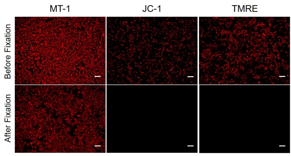
Since mitochondrial membrane potential (MMP) fluctuates according to slight changes of cellular status, extreme care has been needed to obtain repeatable data. In JC-1 dye, which is widely used for measuring the MMP, the fluorescence is lost after a cell has been fixed; therefore, the use of living cells is necessary for the quick measurement of MMP.
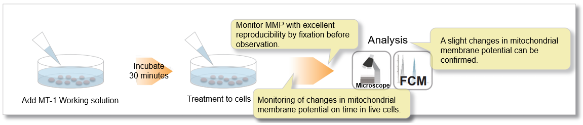
In the meantime, the MT-1 dye, which remains unquenched even in the cells fixed with paraformaldehyde after staining, can be used to conduct a highly repeatable experiment.
② Allow to monitor mitochondrial membrane potential
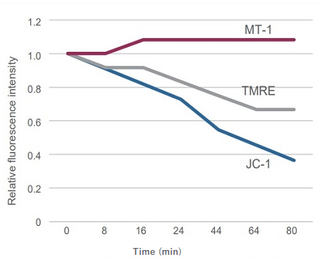
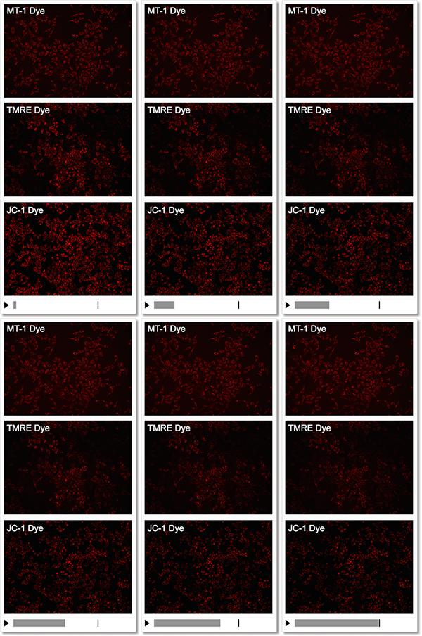
Detection Condition
Ex: 530-560 nm, Em: 570-640 nm
Scale bar: 100 μm
HeLa cells
③ High sensitivity for mitochondrial membrane potential
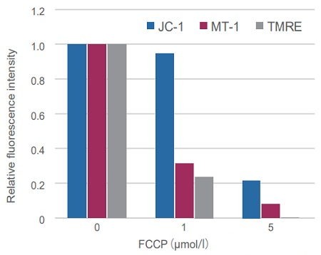
Sometimes it is difficult to detect slight changes of MMP in JC-1 stained mitochondria. In such a case, tetramethylrhodamine ethyl ester (TMRE) was used to monitor MMP. MT-1 can provide equivalent detection sensitivity to TMRE.
Procedure
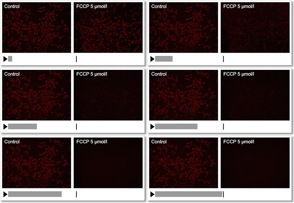
Experimental Example: Depolarization
HeLa cells were treated with a depolarizing agent, carbonyl cyanide-p-trifluoromethoxyphenylhydrazone (FCCP), and changes in mitochondrial membrane potential were observed in a time-lapse imaging with this kit.
As a result, it was confirmed that the mitochondrial membrane potential of the cells treated with FCCP decreased.
Experimental Example: Changes in mitochondrial membrane potential in apoptosis-induced cells
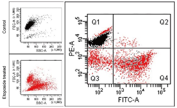
Apoptosis was induced by Etoposide to HL60 cells stained with MT-1 in advance, then co-stained with Annexin V-FITC and analyzed by flow cytometry.
Apoptotic progress and MMP change were confirmed through the fluorescence intensity changes of Annexin V-FITC (an increase in the green fluorescence intensity) and MT-1 (a decrease in the red fluorescence intensity), respectively.
Detection Condition
Annexin V-FITC (green): Ex. 488 nm / Em. 500 – 560 nm
Mitochondria membrane potential (MT-1, red): Ex 488 nm / Em 564-604 nm
Application Data: Simultaneously evaluation of mitochondrial superoxide and membrane potential
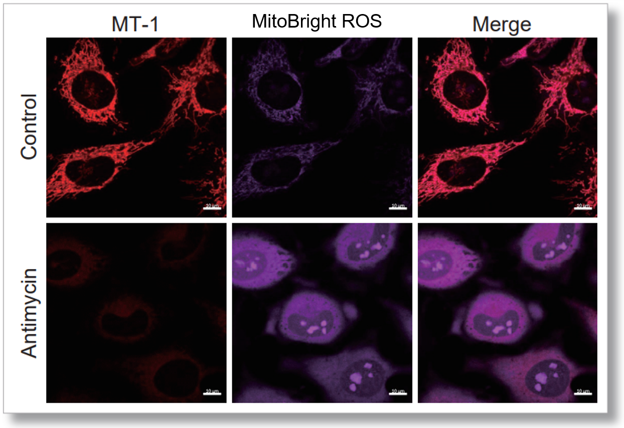
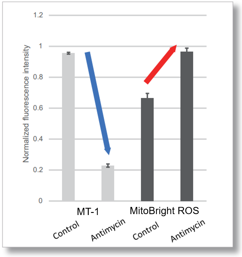
After HeLa cells were washed with HBSS, co-stained with MT-1 MitoMP Detection Kit and mitochondrial superoxide detection dye (MitoBright ROS Deep Red: Code MT16), and the generated mitochondrial ROS and membrane potential were observed simultaneously. As a result, the decrease in mitochondrial membrane potential and the generation of mitochondrial ROS are simultaneously observed.
<General Protocol >


<Imaging Conditions>(Confocal microscope)
MT-1: Ex=561, Em=560-600 nm
MitoBright ROS: Ex=633 nm, Em=640-700 nm
Scale bar: 10 μm


<Examination Conditions>(Plate Reader)Tecan, Infinite M200 Pro
MT-1: Ex=540-550 nm, Em=590-610 nm (Gain=200)
MitoBright ROS: Ex=545-555 nm, Em = 665-685 nm