








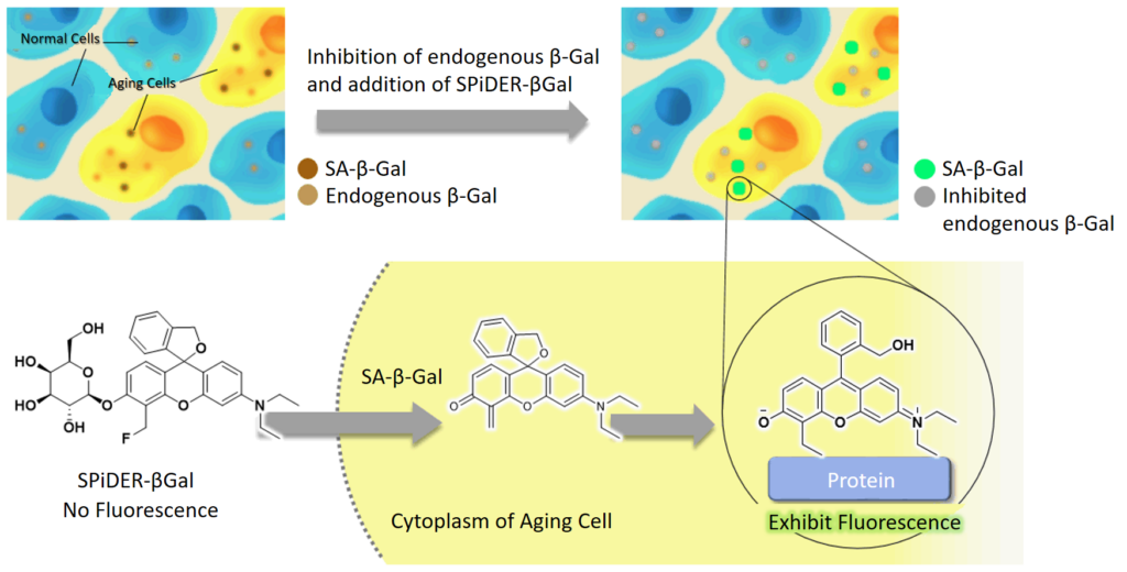
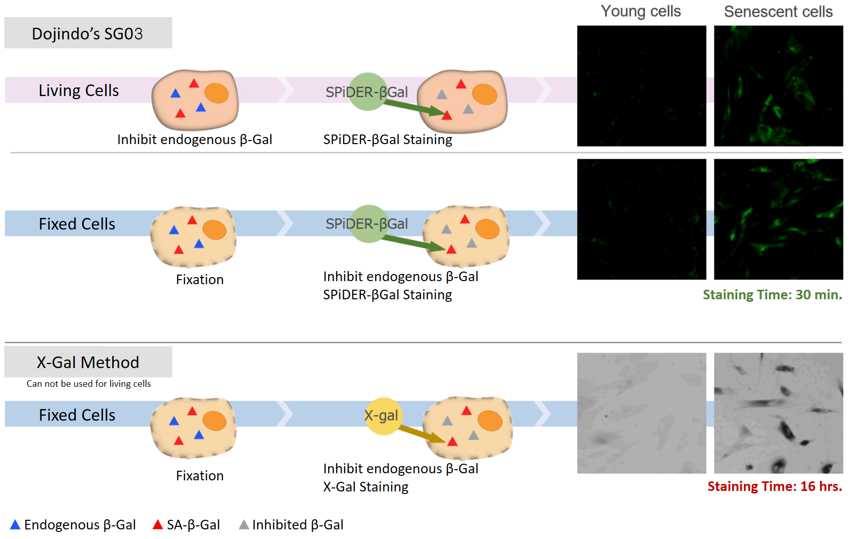
DNA damages of the normal cells are caused by repeated cell division and oxidative stress. Cellular Senescence, a state of irreversible growth arrest, can be triggered in order to prevent DNA-damaged cells from growing. Senescence-associated β-galactosidase (SA-β-gal), which is overexpressed in senescent cells, has been widely used as a marker of cellular senescence. Although X-gal is a well known reagent to detect SA-β-gal, these are following disadvantages: 1) requirement of fixed cells due to the poor cell-permeability, 2) low quantitative capability because of the difficulty of the determination of visual difference between stained cells and not stained cells, 3) requirement of a long time of staining.
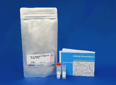
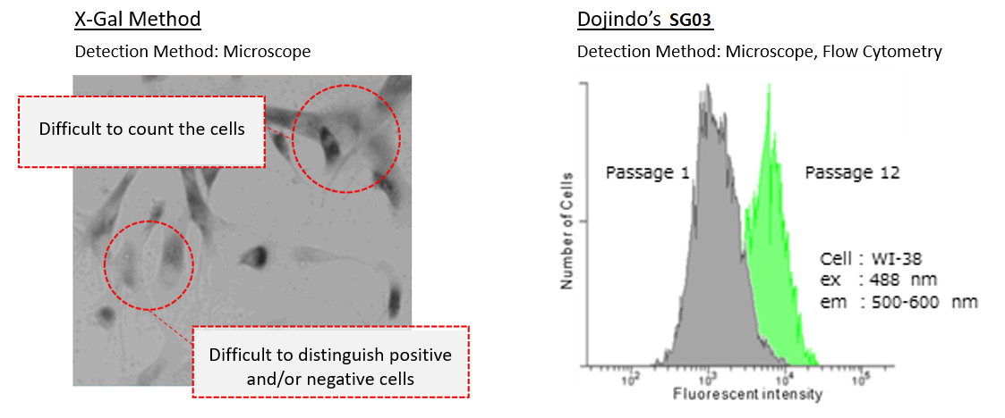
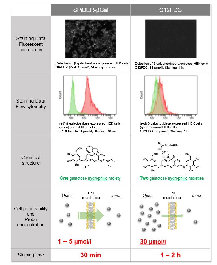
Cellular Senescence Detection Kit – SPiDER-βGal allows to detect SA-β-gal with high sensitivity and ease of use. SPiDER-βGal is a new reagent to detect β-galactosidase which possesses a high cell-permeability and a high retentivity inside cells. SA-β-gal are detected specifically in living cells by using a reagent (Bafilomycin A1) to inhibit endogenous β-galactosidase activity. Therefore, SPiDER-βGal can be applied to quantitative analysis by flow cytometry.
Recent work from Dr. Kim et al. at Mayo Clinic used our Cellular Senescence Detection Kit – SPiDER-βGal to evaluate cellular senescence in endothelial cells. They did staining of SA-βGal in cells in atherosclerotic renal artery stenosis (ARAS) and co-stained the cells with CD31 which is a marker of endothelilal cells. They showed that ARAS + Elamipretide* treatment slightly improved endothelial cell senescence. Unlike commercial available probes used for detection of β-Galactosidase, SPiDER-βGal contained in the kit possesses high intracellular retention. The key feature of this product is that it can be used to co-stain SA-β-Gal and other markers. Our kit is a useful tool for cellular senescence research. *Elamipretide: mitochondria-targeted peptide
For more information on data, please refer to the publication below:
S. R. Kim, A. Eirin, X. Zhang, A. Lerman and L. O. Lerman, “Mitochondrial Protection Partly Mitigates Kidney Cellular Senescence in Swine Atherosclerotic Renal Artery Stenosis.”, Cell. Physiol. Biochem. ., 2019, 52, 617.
Cellular Senescence Analysis Products
| Product Name | Detection | Sample | Dyes / Fluorescence Properties |
|---|---|---|---|
| Cellular Senescence Detection Kit – SPiDER-βGal | Microscopy or FCM | Living / Fixed cells | SPiDER-βGal Ex: 500–540 nm / Em: 530-570 nm |
| Cellular Senescence Detection Kit – SPiDER Blue | Microscopy, FCM or Plate reader | Fixed cells | SPiDER Blue Ex: 350-450 nm / Em: 400-500 nm |
| SPiDER-βGal | Microscopy | Tissue | SPiDER-βGal Ex: 500–540 nm / Em: 530-570 nm |
| Cellular Senescence Plate Assay Kit – SPiDER-βGal | Plate reader | Living cells | SPiDER-βGal Ex: 500–540 nm / Em: 530-570 nm |

Technical info

Difference between X-Gal method and Cellular Senescence Detection Kit – SPiDER-βGal I
Why is Bafilomycin A1 added?
Endogenous β-galactosidase existing in living cells interfere with selective detection of SA-β-Gal. Bafilomycin A1 is an inhibitor of ATPase in lysosome. pH in lysosome is kept neutral by adding Bafilomycin A1. Cellular Senescence Detection Kit – SPiDER-βGal contains Bafilomycin A1 which allows to detect SA-β-Gal selectively. Bafilomycin A1 is utilized for living cell assays only. Bafilomycin A1 is not used in fixed cells because intracellular pH is controlled with the buffer.
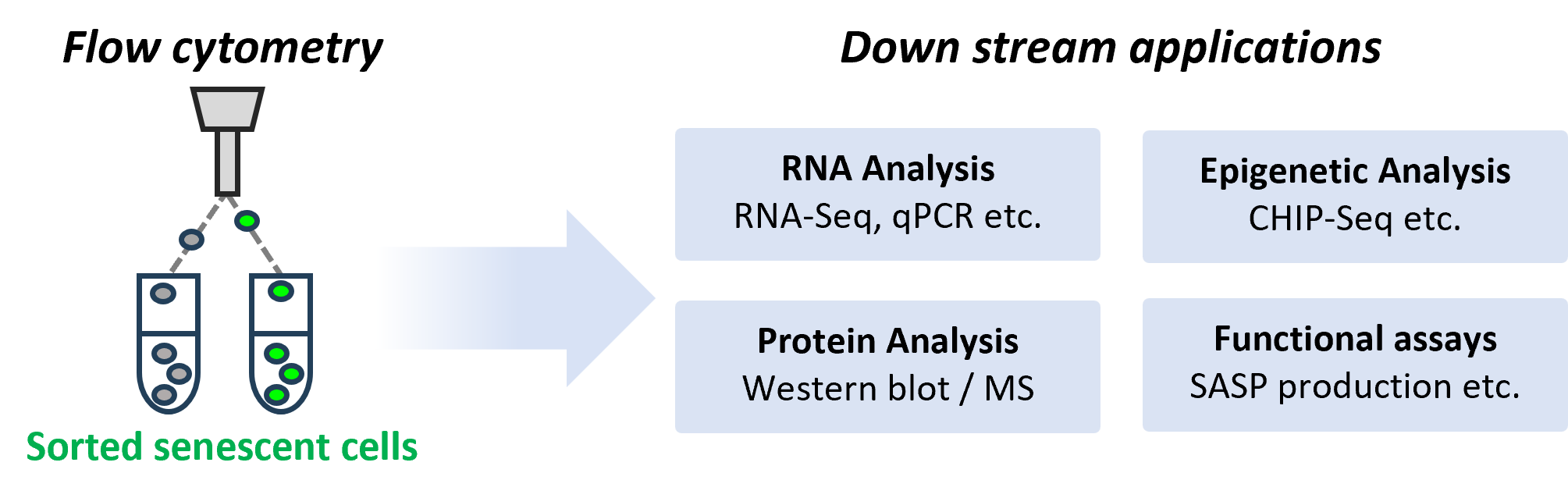
Live-Cell Senescence Detection via Flow Cytometry
Dojindo’s senescence detection kit allows the use of live cells, making post-sorting processes with flow cytometry remarkably simple.
Research Articles
Turano PS, et al., “Age-independent and targetable transcription factor networks regulating CD8⁺ T cell senescence in aging humans.“, Cell Reports, 2026, DOI: 10.1016/j.celrep.2025.116795
Satoru M, et al., “TGF-β in the microenvironment induces a physiologically occurring immune-suppressive senescent state“, Cell Reports, 2023, DOI: 10.1016/j.celrep.2023.112129
Protocol Publication
Pangrazzi L, Martic I, Cavinato M, Weinberger B. “Detecting Senescence in T Cells by Flow Cytometry Using the SA-β-Galactosidase Assay.” In: Gorgoulis VG, Cavinato M, Evangelou K, eds. Oncogene-Induced Senescence. Methods in Molecular Biology, vol. 2906. Humana, New York, NY; 2025:73–81. DOI: 10.1007/978-1-0716-4426-3_5
Co-staining of SA- β-gal and DNA Damage marker in WI-38 cells
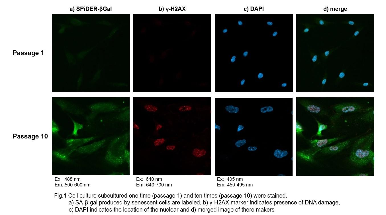
Procedure:
1. Passage 1 and 10 of WI-38 were used. The procedure was followed as the manual within the kit.
2. Add 4% PFA/PBS to the cells and incubate for 15 minutes at room temperature
3. Wash the cells 3 times with PBS
4. Add 0.1% Triton X-100/PBS to cells and incubate for 30 minutes at room temperature
5. Wash the cells 3 times with PBS
6. Add 1% BSA/PBS to the cells and incubate for 1 hour at the room temperature
7. Add anti- γ-H2AX antibody (rabbit) diluted with 1% BSA/PBS to the cells and incubate at 4℃ overnight
8. Wash the cells 3 times with PBS
9. Add Anti- rabbit secondary antibody (Alexa Fluor 647) diluted with 1% BSA/PBS to the cells and incubate at room temperature for 2 hours
10. Wash cells 3 times with PBS
11. Add 2 μg/ml DAPI (code: D523) diluted with PBS to the cells and incubate for 10 minutes at room temperature
12. Wash cells 3 times with PBS and observe under a confocal microscope
Co-staining of SA- β-gal and DNA Damage marker in fixed WI-38 cells
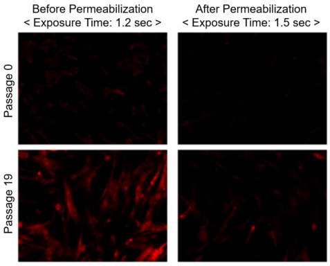
Figure 1. Difference in Fluorescence Intensity of SPIDER-βGal Staining
SPIDER-βGal (red): Ex. 533-548 nm/Em. 570-640 nm
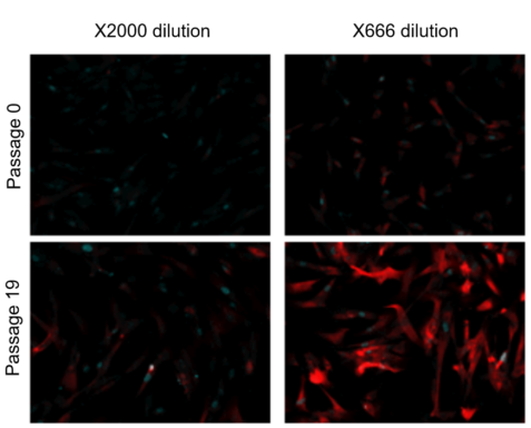
Figure 2. SPIDER-βGal Staining with Different Dilution Ratio
SPIDER-βGal (red): Ex. 533-548 nm /Em. 570-640 nm
γ-H2AX (blue): Ex. 590-650 nm/Em. 668-733 nm
Exposure time: 1.5 sec
Preparation of SPiDER-βGal working solution
Dilute the SPiDER-βGal DMSO stock solution 2,000 times *1 with McIlvaine buffer (pH 6.0).
*1 Fixation and permeablization could leads to lower sensitivity (Figure 1), if you need higher signals,dilute the SPiDER-βGal DMSO stock solution 500 – 1,000 times with the McIlvaine buffer (Figure 2).
Preparation of McIlvaine buffer (pH 6.0)
Mix 0.1 mol/l citric acid solution (3.7 ml) and 0.2 mol/l sodium phosphate solution (6.3 ml). Confirm the pH is 6.0. If the pH is not 6.0, adjust the pH by adding either citric acid solution or sodium phosphate solution. Dilute this buffer 5 times with ultrapure water.
Staining procedure (35 mm dish)
1. Prepare cells on 35 mm dish for assay and culture the dish at 37℃ overnight in a 5% CO2 incubator.
2. Remove the culture medium. Add 2 ml of 4% paraformaldehyde (PFA) /PBS solution to the cells and incubate at room temperature for 3 minutes *2.
*2 Avoid a longer treatment period, which leads to decrease in SA-β-gal activity.
3. Remove the supernatant, and wash the cells 3 times with 2 ml of PBS.
4. Add 2 ml of SPiDER-βGal working solution and incubate at 37℃ for 30 minutes*3.
*3 We recommend not to use a 5% CO2 incubator for fixed cell experiments. If incubation is done in a 5% CO2 incubator, the pH of the buffer may become acidic.
Acidic pH results in higher background from the endogenous β-galactosidase activity and it would be difficult to distinguish between normal cells and senescent cells.
5. After removing the supernatant, wash the cells twice with PBS.
6. Add 0.1% Triton X-100/PBS to cells and incubate for 30 minutes at room temperature.
7. Wash the cells twice with PBS.
8. Add 1% BSA/PBS to the cells and incubate for 1 hour at the room temperature
9. Add anti- γ-H2AX antibody (mouse) diluted with 1% BSA/PBS to the cells and incubate at 4℃ overnight.
10. Wash the cells 3 times with PBS.
11. Add anti- mouse secondary antibody (Cy5) diluted with 1% BSA/PBS to the cells and incubate at room temperature for 1 hour.
12. Wash cells twice with PBS and observe under a fluorescence microscope.
SA-β-gal detection for T cells (floating cells)
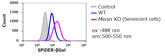
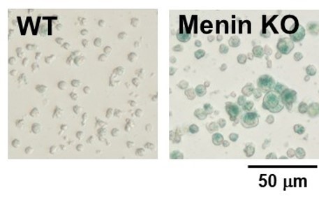
The research group by professor Masakatsu Yamashita at Ehime University Graduate School of Medicine has shown that a protein called Menin controls T cell exhaustion, aging, and maintains normal immune function.
By using this kit, they confirmed that SG03 has the ability to detect induced cell senescence by stimulating TCR (T cell receptor) in the presence of interleukin 2 (IL-II) in naive CD8+ T cells that are knocked out Menin.
Staining Conditions
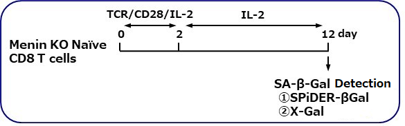
① SPiDER-βGal method
② X-gal method
*Data was kindly provided by Masakatsu Yamashita, at Ehime University Graduate School of Medicine
Quantification with confocal quantitative image cytometer
In the conventional method of X-gal, SA-β-gal-positive cells are counted under microscope and calculate the percent of the senescent cells by compared with total cells. The SA-β-gal-positive cells were stained with this kit and analyzed using confocal quantitative image cytometer CQ1(Yokogawa Electric Corporation).
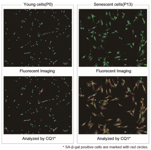
<Imaging Condition> ulture Plate: 96 well plate
Objective Lens: 10 timesEmission Detector
405 nm (Hoechst 33342): Cyan
488 nm (SA-B-gal): Green
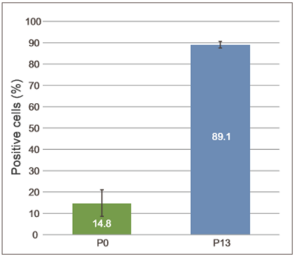
Quantification for SA-B-gal-positive cells
The total cells were stained with DNA staining dye, Hoechst 33342. The SA-β-gal-positive cells and total were counted and calculate the ratio of senescent cells.
The difference of SA-β-gal-positive cells ratio were shown in WI-38 cells depending on the number of passage. The data was quickly analysed with the confocal quantitative image cytometer compared with the manually counting procedure with X-gal staining method.
SA-β-gal detection for tissue
In this published article, SA-β-gal was detected using SPiDER-βGal on tissue samples from a diabetic model mouse.
Note: SPiDER-βGal [Code: SG02] is used in this staining.
After slicing the frozen tissue, it was fixed with 4% paraformaldehyde for 20 minutes at room temperature. Then it was washed with PBS and observed.For details of the experimental operation and data, refer to Reference 2) below.
Properties of SPiDER-βGal
Difference between SPiDER-βGal and comventional reagent (C12FDG)
Excitation and emission spectra
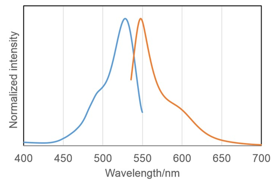
- < Recommended Filter >
Ex: 500-540 nm
Em: 530-570 nm
< Practiced conditions in application data >
Confocal microscopy Ex: 488 nm Em: 500-600nm
Reflected Light Microscopy Ex: 545/25 nm Em: 605/70 nm
Flow Cytometer Ex: 488nm Em: 530/30 nm
Association between cellular senescence and cell cycle
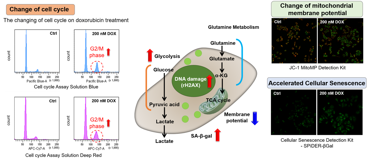
Doxorubicin (DOX) acts to inhibit cell proliferation during G2/M phases of the cell cycle and induces cellular senescence. After adding DOX to A549 cells, higher histogram peaks for the G2/M phase (Cell Cycle Assay Solution Blue and Deep Red), induces cellular senescence (Cellular Senescence Detection Kit – SPiDER-βGal), and the differences in mitochondrial membrane potential (JC-1 MitoMP Detection Kit) were observed.
Markers of Senescent Cells
Senescence Markers
-
・SA-B-Gal (Senescence-Associated Beta-Galactosidase) ・CDKIs (cyclin-dependent kinase inhibitor)
e.g. p16, p21 etc・p53 gene
・SASP (Senescence-Associated Secretory Phenotype)
e.g. IL6, IL8 etc・DDR (DNA damage response)
e.g. y-H2AX, ATM etc・Others
- Multiple markers are utilized to detect cellular senescence. SA-β-Gal is commonly used as a marker of cellular senescence. Cellular Senescence Detection Kit – SPIDER-β-Gal is a suitable kit for imaging and/or quantifying SA-β-Gal.